
Still need help?
Get in touch with the LX.lab team by logging a ticket via ServiceConnect. We'll be in touch shortly.
Log a ticketWant to provide feedback on this resource? Please log in first via the top nav menu.
Learn how Elise Robinson from the Graduate School of Health and the Inclusive Practices Team made X-rays accessible for a student with low vision.
Elise Robinson, physiotherapy lecturer at the Graduate School of Health, sought to improve the visibility of X-rays in her course to better support a student with low vision.
X-rays are used throughout the Canvas modules for 96088 Acute Physiotherapy Care. For instance, they are shown in weekly masterclass slides and simulation classes. The student is expected to analyse a range of X-rays to identify lung and cardiac health, as well as bone or rib injuries, to determine the most effective treatment for the patient.
The student experiences vision loss and finds it difficult to see detailed parts of an x-ray. X-rays are usually in greyscale (black & white), and the lack of contrast between white/grey and grey/black causes accessibility problems for this student. However, the student has the ability to view large size 36-40 text as well as bright colours.
This solution was determined through collaboration with both the student and their Accessibility Consultant. It’s important to note that students with the same disability may have different access needs.
Specific parts of x-rays were edited within Adobe Lightroom to enhance contrast and exposure using the brush tool. This process emphasised the sharpness/bluntness of angles on a chest x-ray to indicate lung health. Arrows and circles were utilised within some x-rays to highlight specific issues. For example, fractured bones and ECG leads. Bright colours such as reds and pinks were employed to add contrast and increase visibility. This enhanced version of the x-ray serves a critical purpose: it aids the student in interpreting what the x-ray report depicts, particularly during clinical placements and as they embark on their professional journey, since they’ve encountered these visuals before.
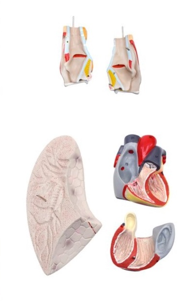
To ensure that the team met the student’s exact access requirements, Accessibility Services engaged in regular consultations with the student, testing each of the samples and providing the team with feedback to improve new iterations. A medical model of the heart and lungs, with the components being detachable, were used alongside these x-rays. This enabled students to gain a clearer understanding of the placement, function, and features of these organs in relation to the ribs, collarbone, and spine.


As you consider the images you use in your teaching, take a moment to reflect on their purpose. Is it necessary to use x-rays, or are you including them for a particular reason? Would a model be a better alternative for communicating the same concept? Ask yourself if the image is intended to convey a concept or if you’re using it as part of your teaching material for a specific instructional purpose.
Do you need to outline or highlight certain areas, or perhaps circle key elements or use arrows to direct attention? Do you need anything labelled? Additionally, think about whether certain parts of the image needs to be brightened or darkened to increase contrast, making it clearer and more visible for your students.
Get in touch with the LX.lab team by logging a ticket via ServiceConnect. We'll be in touch shortly.
Log a ticketWant to provide feedback on this resource? Please log in first via the top nav menu.